Digital diagnostics for the middle ear with endoscopic optical coherence tomography and machine learning |
Medical Need
Multiple conditions of the middle ear are leading to conductive hearing loss, e.g. chronic otitis media, cholesteatoma and otosclerosis, which is regularly investigated by differential diagnosis of several procedures. Current diagnostics cover only single aspects of the pathologies: Otoscopy provides a visual impression and tympanometry the compliance of the tympanic membrane, while audiometry evaluates the overall hearing performance. Nevertheless, all of those tools lack a determination of the leading origin and site of the transmission loss. Accordingly, direct otologic diagnostics enabling the localisation and differentiation for targeted treatment and improved patient outcome are needed.
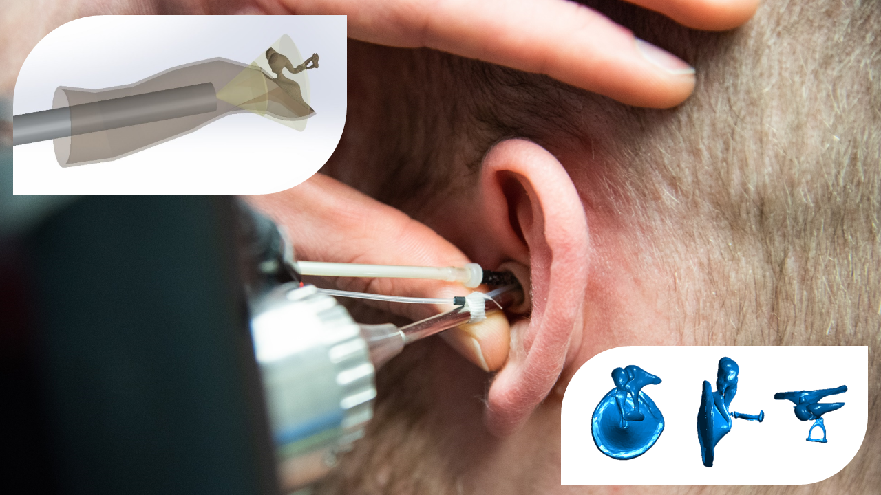
We aim to enable novel digital diagnostics of the middle ear by combining non-invasive optical imaging technology with machine-learning-based image analysis for the immediate and direct examinations of the human organ of hearing, providing the 3D functional assessment of the eardrum and ossicles.
Optical Coherence Tomography, Machine Learning, Middle Ear, Hearing Loss, 3D Imaging, Segmentation, Registration
Morgenstern, J, Schindler, M, Kirsten, L, Golde, J, Bornitz, M, Kemper, M, Koch, E, Zahnert, T, Neudert, M (2020). Endoscopic Optical Coherence Tomography for Evaluation of Success of Tympanoplasty. Otology & Neurotology, 41(7), pp.e901-e905.
Kirsten, L., Schindler, M., Morgenstern, J., Erkkilä, M. T., Golde, J., Walther, J., … & Koch, E. (2018). Endoscopic optical coherence tomography with wide field-of-view for the morphological and functional assessment of the human tympanic membrane. Journal of biomedical optics, 24(3), 031017.
Bodenstedt, S., Rivoir, D., Jenke, A., Wagner, M., Breucha, M., Müller-Stich, B., … & Speidel, S. (2019). Active learning using deep Bayesian networks for surgical workflow analysis. International journal of computer assisted radiology and surgery, 14(6), 1079-1087.
Pfeiffer, M., Riediger, C., Weitz, J., & Speidel, S. (2019). Learning soft tissue behavior of organs for surgical navigation with convolutional neural networks. International journal of computer assisted radiology and surgery, 14(7), 1147-1155.
Reichard, D., Bodenstedt, S., Suwelack, S., Mayer, B., Preukschas, A., Wagner, M., … & Speidel, S. (2015). Intraoperative on-the-fly organ-mosaicking for laparoscopic surgery. Journal of Medical Imaging, 2(4), 045001.
Project team members |
Clinician |
TU Dresden, Faculty of Medicine
University Hospital Dresden, Department of Otorhinolaryngology, Head and Neck Surgery, Ear Research Center Dresden (ERCD)
Project team members |
High-tech |
Abstract |
The aim of the project is to enable novel digital diagnostics for the middle ear by combining innovative optical imaging technology with machine-learning-based image analysis and segmentation. This allows for the first time immediate and direct examinations of the human organ of hearing, providing the 3D functional assessment of the eardrum and ossicles. Multiple conditions of the middle ear are leading to conductive hearing loss, e.g. chronic otitis media, cholesteatoma and otosclerosis, which is regularly investigated by differential diagnosis of several procedures including otoscopy, audiometry, and radiologic diagnostics.
Introducing high-resolution functional and morphologic measurements of the middle ear, endoscopic optical coherence tomography (eOCT) exceeds the capabilities of current diagnostics. However, the evaluation of the complex data sets requires vast expertise and time. A smart digital system based on machine learning and a priori information could provide the treating physician with instant feedback of a patient’s middle ear state. The project will leverage the full diagnostic potential towards a monitoring system in the ENT office and the operating theatre, facilitating the fast diagnosis of conductive hearing loss causes for targeted treatment and improved patient outcome.


