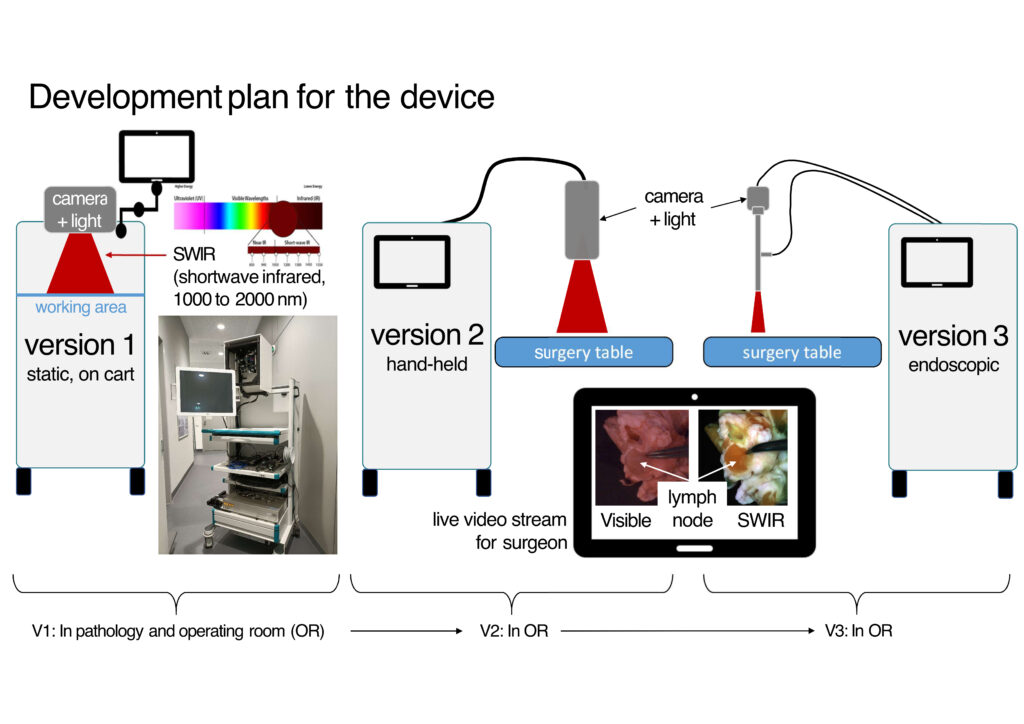
Tumor cells present in lymph nodes are a major predictor of oncological outcome. Accordingly, lymph node detection and resection are mandatory and crucial in oncological surgery. However, identifying lymph nodes is often challenging due to the low contrast with surrounding fatty tissue. Recently, a shortwave infrared (SWIR) imaging approach has been developed to enhance contrast between lymph nodes and fatty tissue. Building on this approach, a SWIR-based lymphatic imager prototype is proposed, initially for testing in pathology labs and ultimately for intraoperative use.
Contrast agent-free imaging of lymph nodes and vessels in the shortwave infrared for surgical oncology
Developing a real-time optical system for lymph tissue detection, enhancing precision and efficiency in oncological surgery
Project Goals
Contrast analysis of lymph nodes and vessels at various SWIR wavelengths
Since lymph nodes/vessels appear darker in the SWIR spectrum due to higher water content, multispectral analysis will determine the optimal wavelength for maximum contrast. Using a NIR-SWIR laser system, preclinical and clinical samples will be examined across wavelengths from 800 to 1600 nm.
Design of an imager for pathology labs
Based on findings from the previous step, a SWIR imaging system will be developed to assist in lymph node detection in the pathology lab. Testing will involve patient samples analyzed by a pathologist and then independently assessed using visible and SWIR imaging to compare detection performance and time efficiency.
Development of intraoperative imaging systems
After successful testing in pathology lab, a wide-field imaging system and an endoscope for intraoperative use will be designed. Ex-vivo testing incl. feedback from surgeons will pave the way for integration into clinical workflows, with plans to prepare for medical device regulatory approval.


