
new publication

What does AI actually learn from tissue images?

Foundation models are increasingly used as the basis for AI systems in computational pathology. Their learned representations can support a wide range of downstream tasks, from tissue characterization to disease classification and biomarker prediction. But strong predictive performance does not reveal which morphological features the AI has actually learned or how it organizes different biological and disease concepts. Researchers from Kather Lab at the EKFZ for Digital Health at TUD Dresden University of Technology, together with pathologists from Mainz and Augsburg, have investigated new ways to make these learned patterns visible. The researchers found that clearly distinct tissues were represented through morphological patterns that pathologists could also recognize. By contrast, cancer types that look similar under the microscope also showed greater overlap in the AI model’s representations. The visualization framework could therefore help researchers examine what pathology AI has learned – and where distinctions between disease concepts become less clear. The study was published in Cell Reports Medicine.

Marco Gustav*, Fabian Wolf*, Christina Glasner*, Nic G. Reitsam, Stefan Schulz, Kira Aschenbroich, Bruno Märkl, Sebastian Foersch#, Jakob Nikolas Kather#: Class Visualizations and Activation Atlases for Computational Pathology, Cell Reports Medicine, 2026.

(* equal first authorship | # equal last authorship)

Visualization approaches

Foundation models are trained on extensive image collections and can subsequently be adapted to different diagnostic or research tasks. Yet even when such systems perform well, what they have learned often remains hidden within their internal representations and is difficult to interpret. To make these learned patterns more accessible, the researchers combined two complementary visualization approaches: class visualizations and activation atlases.
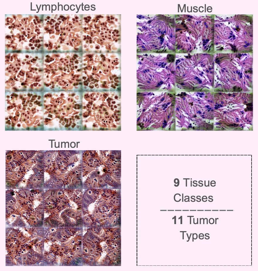
Class visualizations are synthetic images representing patterns that strongly activate a particular tissue or cancer category in the AI model. They provide a glimpse of the features the model associates with concepts such as fat tissue, lymphocytes, or tumor tissue.
Class visualization - squares showing different tissue types like Lymphocytes, muscle an tumor

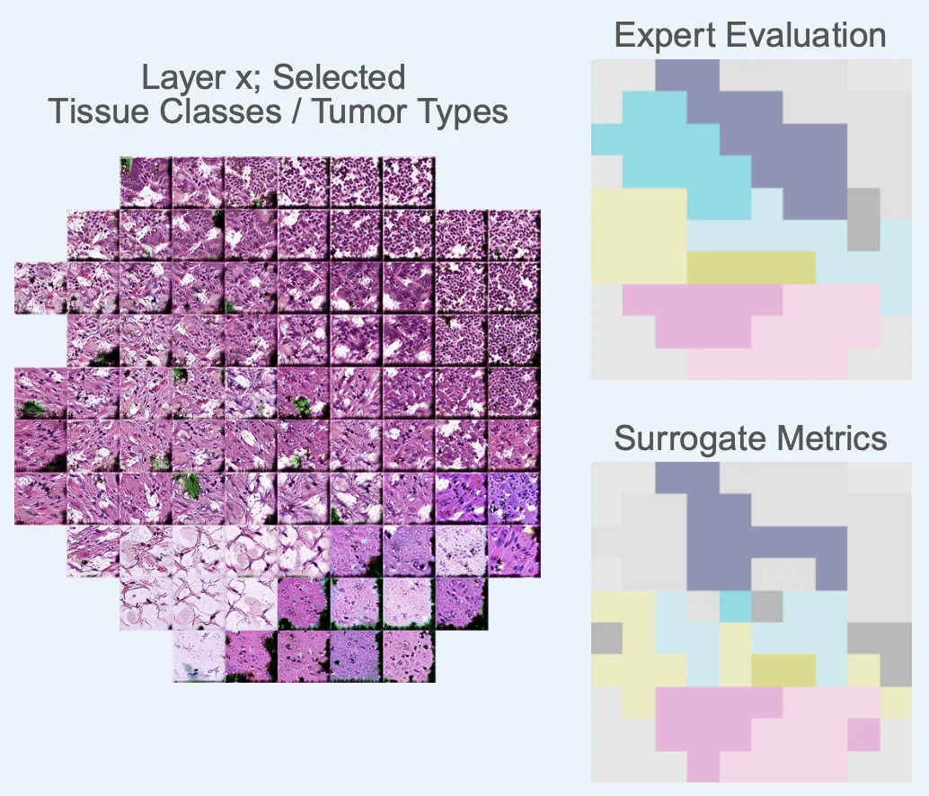
Activation atlases take a broader perspective. Instead of focusing on a single category, they create a visual map of how the AI organizes different tissue patterns internally – showing which concepts are represented separately and where they begin to overlap.

Activation Atlases showing tissue classes and tumor types

“The important question is not only whether an AI model gives the right answer, but which morphological features and patterns it relies on,” says Marco Gustav, PhD, shared first author of the study and postdoctoral researcher at the EKFZ for Digital Health at TU Dresden. “These visualizations allow pathologists to examine whether the model relies on recognizable morphological patterns, how clearly it distinguishes between different tissue or disease concepts, and which patterns these concepts share. In the future, this could work both ways: we may not only learn more about which morphological patterns AI models rely on, but AI could also point us towards patterns that deserve closer scientific investigation.”


Recognizable patterns, but less clear distinctions


Four pathologists independently evaluated real tissue images and the AI-generated visualizations without knowing the intended labels. The researchers tested the approach on colorectal tissue and several cancer types, ranging from relatively distinct tissue categories to more challenging tumor classifications. The picture became less clear as the diagnostic task became more complex. Adenocarcinomas from different organs can share very similar histomorphological features, even though they arise in different tissues. Their AI representations also showed greater overlap and were harder for pathologists to distinguish. This effect increased for more narrowly defined cancer subtypes. Activation atlases showed the same overall pattern: broader tissue and cancer categories formed more coherent regions, while morphologically similar categories increasingly overlapped. The researchers also found that the organization of information changed across the neural network: earlier layers were dominated by more basic visual features, while deeper layers showed increasingly specialized representations.

The visualization methods do not explain exactly why an AI system makes an individual prediction, but they provide a way to inspect how a model represents morphological concepts and where these concepts overlap. This could help researchers determine whether an AI model for pathology has learned morphologically meaningful features or whether it relies on potential artifacts or dataset-specific shortcuts. It could also help them compare AI representations with expert pathological knowledge. Future studies will need to test the approach across other foundation models, diseases, and datasets. To support this, the code and an interactive visualization tool are openly available. The authors aim to encourage and enable researchers and pathologists to use the framework to explore their own models, datasets, and scientific questions.

Share this Post

More News

On-premise medical AI agent for reliable clinical decision support
Skip to content